Anterior vs Posterior Placenta: What It Means
<p>The main difference between anterior and posterior placenta is location. An anterior placenta attaches to the front uterine wall, possibly muffling fetal movements. A posterior placenta attaches…
- Published
- Written by
- wib-seo
- Reading time
- 6 minutes
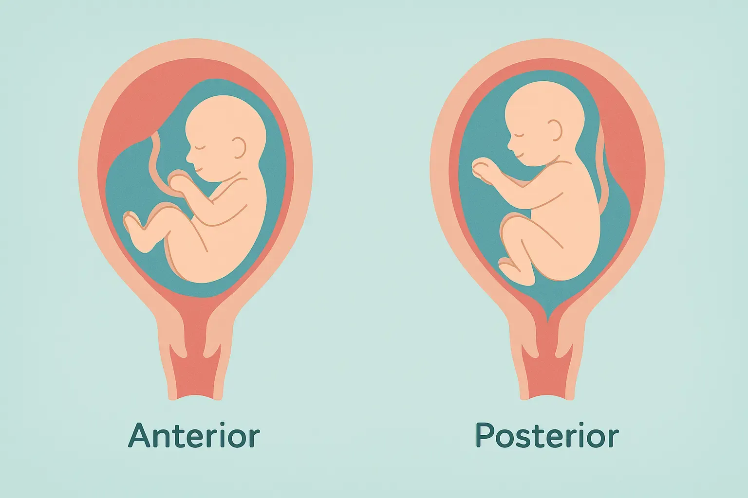
The main difference between anterior and posterior placenta is location. An anterior placenta attaches to the front uterine wall, possibly muffling fetal movements. A posterior placenta attaches to the back wall, often making kicks easier to feel. Both positions are normal and rarely affect pregnancy outcomes.
Key Takeaways
- Anterior vs posterior placenta positions are both completely normal variations that don’t increase pregnancy risks
- Anterior placenta may cushion fetal movements, making kicks feel softer or take longer to notice
- Placental position is routinely noted during ultrasounds but rarely affects delivery plans unless other complications are present
When you receive your ultrasound report and see “anterior placenta” or “posterior placenta” mentioned, it’s natural to wonder what this means for you and your baby. These terms simply describe where your placenta has attached inside your uterus – information that helps your healthcare team monitor your pregnancy but usually doesn’t change your care plan.
Understanding anterior vs posterior placenta positions can help ease any concerns and give you insight into what to expect as your pregnancy progresses, particularly regarding fetal movement patterns and routine monitoring.
What “Placenta Position” Means
Placenta position refers to where your placenta has implanted and grown along the walls of your uterus. Think of your uterus as having different walls – front, back, top, and sides – where the placenta can attach.
The anterior wall is the front wall of your uterus, closest to your abdominal wall and belly button. When your placenta attaches here, it’s called an anterior placenta.
The posterior wall is the back wall of your uterus, closer to your spine. A placenta attached here is called a posterior placenta.
Both positions are completely normal and occur in roughly equal frequencies. Your placenta’s position is determined by where your fertilized embryo initially implanted, which happens randomly during early pregnancy development.
The placenta can also attach to the sides (lateral) or top (fundal) of the uterus, though anterior and posterior are the most commonly discussed positions.
Anterior Placenta — What to Expect
An anterior placenta sits between your baby and your abdominal wall, essentially acting as a cushion. This positioning creates some unique experiences during pregnancy.
Fetal Movement Patterns
You may notice fetal movements slightly later than women with posterior placentas, typically around 18-22 weeks instead of 16-20 weeks for first pregnancies. The placenta doesn’t prevent movement detection but can muffle the sensation.
Kicks and movements may feel softer or more cushioned throughout pregnancy. Some women describe feeling movements more toward their sides rather than directly in front.
Movement counts remain important – you should still feel regular daily movement once patterns are established, usually by 24-28 weeks.
Monitoring Considerations
Early Doppler detection of your baby’s heartbeat might take a few extra weeks, as the placenta can interfere with sound transmission. This is completely normal and not concerning.
Ultrasound imaging may require different angles or approaches, but skilled technicians routinely work with anterior placentas without difficulty.
Routine prenatal care continues exactly as normal – an anterior placenta alone doesn’t increase any pregnancy risks or require additional monitoring.
Posterior Placenta — What to Expect
A posterior placenta attaches to the back wall of your uterus, leaving the front area more open for direct contact with your baby.
Fetal Movement Patterns
Earlier movement detection is common, sometimes as early as 16-18 weeks in first pregnancies and even earlier in subsequent pregnancies.
Stronger, more pronounced movements are typically felt since there’s less cushioning between baby and your abdominal wall.
Kicks may feel sharper or more defined, especially as your baby grows and movements become stronger.
Monitoring Advantages
Doppler detection of fetal heartbeat often happens earlier and more easily during routine appointments.
Ultrasound imaging typically provides clearer views of your baby since the placenta isn’t blocking the front access.
External monitoring during labor may be more straightforward, though modern equipment works well regardless of placental position.
How and When Position Is Determined
Timing of detection usually occurs during your anatomy scan around 18-22 weeks of pregnancy, though placental position may be noted earlier during routine ultrasounds.
Assessment Methods
Ultrasound technology is the standard method for determining placental position. Technicians can clearly visualize where the placenta has attached and note its relationship to your cervix.
Multiple views during ultrasounds ensure accurate assessment from different angles, providing comprehensive information about placental location and health.
For point-of-care checks outside the hospital, clinicians increasingly use a Portable Ultrasound Machine to confirm placental location and optimize views during routine scans.
Position “Changes” During Pregnancy
Apparent movement of the placenta sometimes occurs as your pregnancy progresses, but the placenta doesn’t actually move. As your uterus grows and stretches, the relative position can appear different.
Growing uterus changes the proportions and angles, which may make an anterior placenta appear less prominent or a posterior placenta seem to shift position slightly.
Final positioning is typically confirmed during third-trimester ultrasounds if needed for delivery planning.
Labor and Delivery Considerations
Placental position alone – whether anterior or posterior – typically has no impact on your delivery method or birth plan. Both positions are compatible with vaginal delivery and don’t increase cesarean section likelihood.
Important Distinctions
Anterior vs posterior position is completely different from conditions that do affect delivery:
Placenta previa occurs when the placenta covers the cervix, potentially requiring cesarean delivery. This is about proximity to the cervix, not front-versus-back positioning.
Low-lying placenta refers to placental position near the cervix and is monitored separately from anterior/posterior classifications.
Delivery Planning
Standard labor management applies regardless of placental position. Your healthcare team notes the position for reference but typically doesn’t alter birth plans based on this information alone.
Procedure considerations may involve placental location for certain interventions, but this is managed by your medical team without requiring changes to your birth preferences.
Pain management options remain the same whether you have an anterior or posterior placenta.
When to Contact Your Provider
While placental position itself rarely causes complications, certain symptoms always warrant medical attention:
Vaginal bleeding at any point during pregnancy, especially if accompanied by cramping or pain.
Severe abdominal pain that doesn’t resolve with rest or position changes.
Fluid leakage that might indicate rupture of membranes, regardless of your gestational age.
Markedly decreased fetal movement after you’ve established your baby’s normal pattern, usually after 28 weeks.
Concerns after ultrasounds or questions about any findings mentioned in your reports.
Trust your instincts – if something feels wrong or different, it’s always appropriate to contact your healthcare provider for guidance.
Conclusion
Understanding anterior vs posterior placenta positions can help you feel more informed about your pregnancy and what to expect regarding fetal movement patterns and routine monitoring. Both positions are completely normal variations that don’t affect your baby’s health or development.
The key takeaway is that placental position is simply anatomical information that helps your healthcare team provide optimal care. Whether anterior or posterior, your placenta is doing its vital job of nourishing your baby throughout pregnancy.
Focus on establishing healthy pregnancy habits – proper nutrition, regular prenatal appointments, and monitoring fetal movements according to your provider’s guidance. These factors matter far more than placental position for pregnancy outcomes.
If you have questions about your specific placental position or any concerns about fetal movement patterns, don’t hesitate to discuss them during your next prenatal appointment.
Frequently Asked Questions
Can placenta position change later in pregnancy?
The placenta stays attached in the same location, but its relative position may appear different as your uterus grows and stretches during pregnancy.
Does placenta position affect C-section likelihood?
No, anterior or posterior placental position alone doesn’t increase cesarean delivery likelihood – only specific complications like placenta previa affect delivery method.
Is anterior placenta the same as placenta previa?
No, anterior placenta refers to front wall attachment while placenta previa means the placenta covers the cervix – these are completely different conditions.