Intrauterine Pregnancy: Definition, Diagnosis, and Stages
Key Takeaways Finding out you’re pregnant brings excitement and questions. One of the first things healthcare providers confirm is that your pregnancy is developing in the right location—inside your…
- Published
- Written by
- Women In Balance Team
- Reading time
- 6 minutes
Key Takeaways
- Intrauterine pregnancy (IUP) occurs when a fertilized egg successfully implants in the uterine lining—the normal, healthy location for pregnancy development.
- Transvaginal ultrasound is the gold standard for confirming intrauterine pregnancy, showing a gestational sac with yolk sac or fetal pole, typically visible at 4.5-5 weeks gestational age.
- Early diagnosis distinguishes normal intrauterine pregnancy from ectopic pregnancy (abnormal implantation), ensuring appropriate prenatal care and monitoring throughout the 40-week pregnancy journey.
Finding out you’re pregnant brings excitement and questions. One of the first things healthcare providers confirm is that your pregnancy is developing in the right location—inside your uterus. Intrauterine pregnancy is the medical term for when a fertilized egg implants and grows in its correct location within the uterine lining. This is the normal, healthy scenario that leads to a full-term pregnancy and delivery. Learning what intrauterine pregnancy means, how it’s confirmed through ultrasound, and what happens during early pregnancy helps expectant mothers navigate the crucial first weeks with confidence. Transvaginal ultrasound is the only way to definitively confirm that implantation occurred in the uterus rather than elsewhere, making early prenatal care essential for pregnancy health and safety.
What Is Intrauterine Pregnancy?
Intrauterine pregnancy occurs when a fertilized egg successfully travels down the fallopian tube, implants in the uterine lining (endometrium), and begins developing into an embryo, then fetus, and eventually a baby. This represents normal pregnancy implantation and is the foundation for a healthy three-trimester pregnancy journey lasting approximately 40 weeks or 280 days from the first day of your last menstrual period.
The process begins with fertilization, typically in the fallopian tube, followed by the fertilized egg traveling to the uterus over several days. Implantation occurs when the fertilized egg embeds itself into the thick, nutrient-rich uterine lining that has prepared for potential pregnancy. Once implantation succeeds, the pregnancy continues developing within the protected environment of the uterus.
Intrauterine pregnancy differs from ectopic pregnancy, where implantation occurs in an abnormal location such as the fallopian tube, ovary, cervix, or abdominal cavity. Ectopic pregnancies cannot develop normally and pose serious health risks, making early confirmation of intrauterine location critical for maternal safety and appropriate care.
How Intrauterine Pregnancy Is Diagnosed
Confirming intrauterine pregnancy requires transvaginal ultrasound imaging. Home pregnancy tests detect pregnancy through hormone levels but cannot determine location—only medical imaging can confirm the pregnancy is developing in the uterus.
Ultrasound confirmation
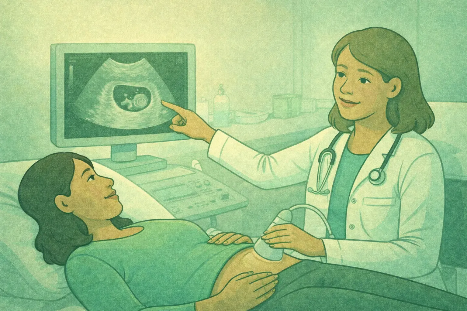
Transvaginal ultrasound is the gold standard for confirming intrauterine pregnancy. This imaging technique uses a probe inserted into the vagina to obtain clear, detailed images of the uterus and early pregnancy structures. Transvaginal ultrasound can visualize pregnancy earlier and more clearly than transabdominal ultrasound.
An intrauterine pregnancy is definitively confirmed when ultrasound shows a gestational sac containing either a yolk sac or fetal pole within the uterus, according to the Society of Radiologists in Ultrasound consensus guidelines. The gestational sac—a fluid-filled structure surrounding the embryo—becomes visible as early as 4.5 to 5 weeks gestational age and is 97.6% specific for diagnosing intrauterine pregnancy, according to research published in StatPearls. However, seeing an empty gestational sac alone isn’t sufficient confirmation, as 10-20% of ectopic pregnancies can produce pseudogestational sacs (fluid collections mimicking early pregnancy).
Key ultrasound findings confirming IUP:
- Gestational sac within the uterine cavity
- Yolk sac visible inside the gestational sac (usually around 5-6 weeks)
- Fetal pole (early embryo) developing within the gestational sac
- Cardiac activity (heartbeat) detected, typically around 6 weeks gestational age
- Appropriate growth corresponding to gestational age
Ultrasound has 92-100% specificity for confirming intrauterine pregnancy when both gestational sac and yolk sac or fetal pole are visualized, according to research from the American College of Emergency Physicians.
Stages of Intrauterine Pregnancy
Pregnancy is measured in weeks and divided into three trimesters, each lasting approximately 13 weeks. Full-term pregnancy is 40 weeks from the first day of your last menstrual period.
First Trimester (Weeks 1-12)
The first trimester is crucial for establishing pregnancy and early development. During the first two weeks, you’re not yet pregnant—fertilization typically occurs around week 2. Implantation happens during week 3-4, and the gestational sac becomes visible on ultrasound around weeks 4.5-5.
Major developments include formation of the neural tube (future brain and spinal cord), development of the heart (which begins beating around 6 weeks), formation of limb buds, and establishment of basic organ systems. The embryo grows from a tiny cluster of cells to approximately 2-3 inches long by the end of the first trimester.
Second Trimester (Weeks 13-26)
The second trimester focuses on growth and organ maturation. Many women find this the most comfortable trimester with decreased nausea and increased energy. The fetus grows rapidly, developing facial features, fingerprints, and the ability to hear. Mothers typically feel fetal movements (quickening) between 16-20 weeks. By the end of the second trimester, the fetus weighs approximately 2 pounds and measures about 14 inches long.
Third Trimester (Weeks 27-40)
The third trimester prepares the baby for life outside the uterus. The fetus gains significant weight, lungs mature for breathing, brain development accelerates, and the baby assumes the head-down position for birth. Regular prenatal visits monitor fetal position, growth, and maternal health as delivery approaches.
Monitoring Early Intrauterine Pregnancy
Once intrauterine pregnancy is confirmed, regular prenatal care monitors pregnancy progression and maternal health.
Early pregnancy monitoring includes:
- Follow-up ultrasounds to confirm fetal cardiac activity and appropriate growth
- Blood tests to monitor pregnancy hormone levels and assess overall health
- Prenatal vitamin supplementation, especially folic acid
- Additional blood tests for blood type, anemia, infections, and genetic screening options
- Lifestyle counseling regarding nutrition, exercise, and substance avoidance
- Discussion of pregnancy risks and warning signs requiring immediate attention
Women with confirmed intrauterine pregnancy should contact healthcare providers if experiencing heavy vaginal bleeding, severe abdominal pain, persistent vomiting, high fever, or other concerning symptoms.
Risks and Complications
While most intrauterine pregnancies progress normally, potential complications can occur:
Miscarriage: Early pregnancy loss, occurring in approximately 10% of clinically recognized pregnancies according to data from the American College of Obstetricians and Gynecologists, most commonly in the first trimester.
Blighted Ovum: Empty gestational sac without developing embryo, diagnosed when mean sac diameter exceeds 25mm without visible embryo.
Molar Pregnancy: Rare condition where abnormal tissue grows instead of normal embryo, requiring prompt treatment.
Preterm Labor: Labor beginning before 37 weeks, requiring medical intervention to delay delivery when possible.
Conclusion
Intrauterine pregnancy represents the normal, healthy implantation of a fertilized egg within the uterus, setting the foundation for fetal development through three trimesters to delivery. Early confirmation through transvaginal ultrasound ensures appropriate prenatal care and rules out ectopic pregnancy requiring immediate intervention.
Knowledge of the stages, ultrasound findings, and monitoring process empowers expectant mothers to actively participate in their prenatal care. With regular medical supervision and healthy lifestyle choices, most intrauterine pregnancies progress successfully to delivery of healthy babies.
Frequently Asked Questions
Can stress affect intrauterine pregnancy?
Moderate stress typically doesn’t harm pregnancy, but chronic severe stress may increase preterm birth and low birth weight risk, according to research published in Developmental Review. Manage stress through rest, support, and relaxation. Discuss overwhelming stress with healthcare providers.
What to avoid during intrauterine pregnancy?
Avoid alcohol, tobacco, drugs, raw meats, unpasteurized dairy, high-mercury fish, and cat litter. Limit caffeine and consult providers before taking any medication during pregnancy.
What are the three types of pregnancies?
The three types are intrauterine pregnancy (normal uterine implantation), ectopic pregnancy (implantation outside uterus), and heterotopic pregnancy (simultaneous intrauterine and ectopic implantation).